Overview

Proven Technology for Diagnostic Excellence

Proven technology for improved workflow efficiency and patient care are set to define digital pathology and increase overall adoption. With the call for more rigorous controls, manufacturers of digital pathology devices are being held to the highest standards of quality, security, and patient safety.

The Aperio GT 450 DX uses innovative color calibration technology to output consistent high-quality images enabling on-time, on-screen diagnosis. Aperio GT 450 DX is an automated, high capacity digital pathology slide scanner that offers exceptional image quality, rapid delivery of cases and high-performance streamlined digital viewing built with the pathologist in mind.

  • Improve case turnaround with no touch scanning and continuous loading of the racks directly from the HistoCore SPECTRA Workstation (Stainer & Coverslipper)
  • Increase IT security and control with a dedicated Scanner Administrator Manager DX (SAM DX) server and software that allows you to set up and monitor multiple Aperio GT 450 DX scanners at a time. No more single workstations for each scanner
  • Ensure excellent image quality with Leica optics
Contact Us to get a quote. Call +1 312 565 6737 to speak with a representative.

For In Vitro Diagnostic Use
FDA 510(k) and CE Marked under the IVDR


Features

Confidence in Diagnosis for the Pathologist
Security and Interoperability for IT Professionals
Speed to Quality for Lab Managers and Histotechnicians

Image Quality Tested by Pathologists for Pathologists

(for diagnostic review use Aperio Webviewer DX)

Image
Mitotic Events (Breast)
Image
Intercellular Bridges
Image
Chromatin
Chromatin
Image
Nucleoli
Nucleoli

Precision, Potential to Scale, and Peace of Mind

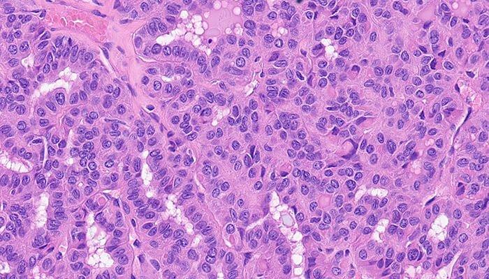
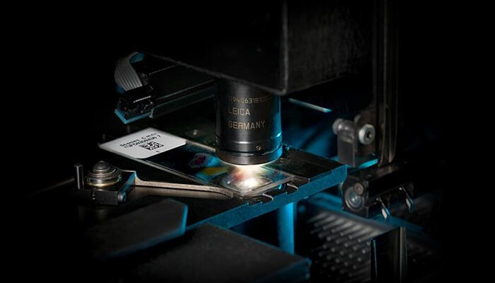
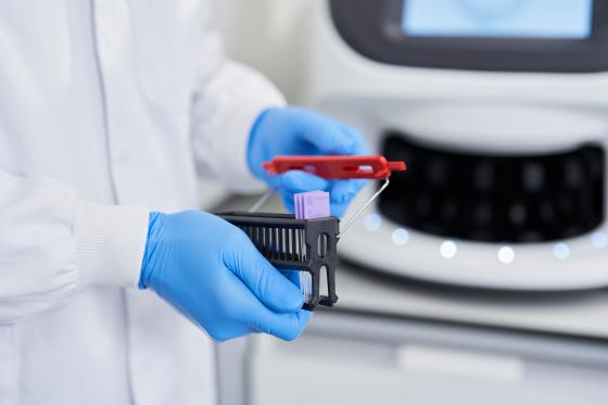
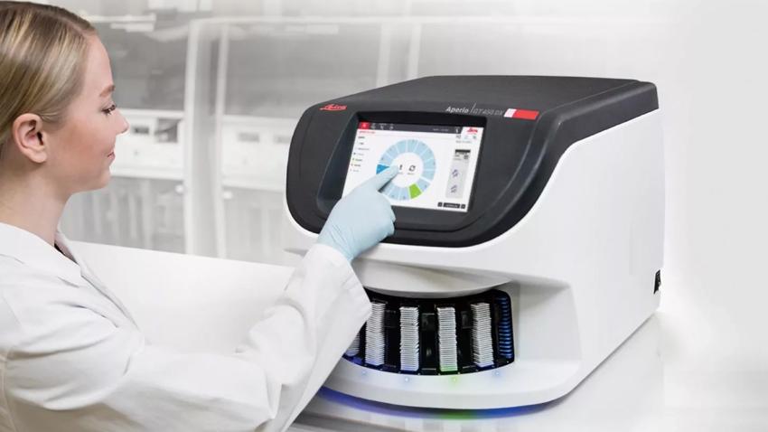
Digital pathology scan speeds have not always been fast enough at 40x magnification to keep up with high-volume scanning (120k+ slides per year). Real-Time Focusing (RTF)** offers a potential solution to this problem. It’s a novel method to capture a digital image that combines an imaging line sensor and a focusing line sensor.

  1. While scanning, the focusing line sensor receives focusing data of the tissue.
  2. Proprietary algorithms determine the best focus value.
  3. Then, each best focus value is treated like a control loop with the mechanics, feeding the best focus value to the objective height on the fly.
  4. The positioning adjustments of the objective are performed continuously (in real time) in order to enable the imaging line sensor to capture the image at its best possible focus. Hence, real time focusing.

**US Patent no. 9,841,590

Image
Cellular Groves (Thyroid)

Excellent Image Quality with Custom-Made Leica Objective

The high-performance objectives included with the Aperio GT 450 DX is specifically designed to maximise field of view for high speed digital pathology scanning. Most objectives are designed similarly to the human eye: round, which focuses light in the center and limits field of view. The objectives on the Aperio GT 450 DX includes an extra-wide flat field correction that enables a much larger field of view (1mm) that can accommodate extremely large digital images for fast scans and 0.26 μm/pixel resolution at 40x magnification.

Image
Excellent Image Quality with Custom-Made Leica Objective

Specifications

Imaging, Capacity & Operations
Dimensions & Weight
IT Requirements & Integration
Security Features
Slides, Coverslips & Labels

Downloads

View all available downloads below

Similar Products

HistoCore SPECTRA Workstation

HistoCore SPECTRA Automated Slide Staining Workstation

The highly automated workstation solution to manage increasing workloads and high throughput demands. Specifically designed to deliver timely and consistent results from slide 1 – 1600.

Knowledge Hub

From tissue processing to Digital Pathology, our solutions empower labs with efficiency, precision, and reliability.

top consideration when buying a scanner
Top Considerations When Buying a Digital Pathology Scanner

Are you thinking about purchasing a digital pathology scanner? We offer some tips for things to consider during the buying process.

4-tips-for-digital-pathology-slide-scanning-640x480
Four Tips for Digital Pathology Slide Scanning

Do you want to get better digital images from your glass slides? Here are some tips to help you get the best out of your digital pathology line scanner: